Vibrio fischeri
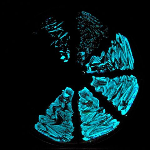
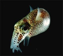
Vibrio fischeri is a gram negative bioluminescent bacterium shaped like a rod that can be found throughout the world’s oceans, but in highest concentration in sub-tropical waters. The cell wall of Vibrio fischeri is made up of polysaccharide and protein. The outer layer of the cell wall contains porins, which are pore like structures for a specific type of molecule. Vibrio fischeri propels itself using a tail called a flagellum. The bacterium is heterotrophic, and feeds off of dead, decaying matter. Although Vibrio can be found in a free planktonic, drifting state, it only produces light while in symbiosis with marine animals, where there is a high enough concentration of bacteria. An animal that hosts Vibrio is the bob tailed squid, Euprmna scolopes, in the form of a mutualistic symbiosis. When the squid is born, it contains none of the bacteria; rather it is acquired throughout the time averaging around a month. At this hatchling stage, the squid has several adaptations to help acquire the sufficient population of bacteria, such as ciliated tentacles or arms, that wave sea water inside of them The bacteria is held within a specialized organ known as the light organ, where the population of bacteria is grown to the point in which it can produce light. After this point, the squid ejects approximately ninety percent of the bacteria each morning in a process called venting, to give the present bacteria area to grow, and it is believed that the ejection may take place as an attempt to provide hatchlings with a great amount of bacteria. Vibrio produced light in a circadian rhythm, meaning it produces more light at night time than during the day. In this symbiosis, Vibrio is provided with food and shelter within the squid, and in return, produces enough light from the organ to provide the squid with the camouflage of the waters surface, and is also used to attract prey. The bacterium emits a blue-green light at the frequency of 490nm (nanometers), this is significant because the spectrophotometer will need to be adjusted to the proper wavelength to provide an accurate reading. The salinity in which vibrio is commonly found is 30ppt, and this measurement will be used as the control in the experiment. Vibrio fischeri follows the equation for bioluminescence: ATP (energy)+Luciferin (a substrate produced by the bacteria)+Luciferase (an enzyme that catalyzes the reaction)+oxygen=>Light. In simple form: ATP+Luciferin+Luciferase+Oxygen=> Light.
Bioluminescence
Bioluminescence is the ability for an organism to naturally produce light. Bioluminescence is commonly used to stun prey, provide camouflage (in Vibrio’s case), or attract other organisms to their location, being mates or prey. Bioluminescence is mostly found in the bathypelagic, abyssopelagic and hedalpelagic zones in the ocean, where the only light present is naturally produced, due to the extreme depth. Nearly every organism able to survive in the hedalpelagic zone is remotely bioluminescent. Bioluminescence is an accurate response indicating an enzyme’s efficiency. If the enzyme is functioning, the bioluminescent organism will emit light. The efficiency can be measured in the quantity of light emitted. For instance if more light is produced, but under both circumstances, light is produced, it shows that although the enzyme can function in both environments, it excels under different conditions and is less effective in another. The substrate that causes organic light to be emitted by an organism is luciferin, and the enzyme that catalyzes all bioluminescent reactions is luciferase. There are two main types of bioluminescent light emissions: A pulsing light and an unchanged light. Pulsing lights will emit light in bursts, where there will be times that no light is emitted and a cap where light is brighter than the rise or the decline. In unchanged light, the organism will always be producing light if the enzymes are functioning properly. The light emitted by a bioluminescent organism often varies from another. For instance, the bacteria being used in the study emits a blue-green light, as some other organisms such as the firefly emits yellow light, and the crustacean sea firefly emits a blue light.
Symbiosis
A symbiotic relationship is a relationship in which one, both or neither organism benefits. The symbiotic relationship relevant to this experiment is between Vibrio (a bioluminescent bacterium) and Euprymna scolopes. In this relationship, both organisms benefit, so it is a mutualistic relationship. There are five types of symbiotic relationships: Mutualism (both organisms benefit), Commensalism (one organism benefits, one unharmed), Parasitism (one species benefits, other suffers), competition (both organisms suffer) and neutralism (both organisms are unaffected. Vibrio’s relationship is mutualistic because the bacteria’s light production provides the bob tailed squid with camouflage, and the bacteria are provided with shelter and food. When the squid is hatched, it contains no symbiots, but rather acquires them throughout time. The hatchling is born with adaptations that make it easier to accumulate the necessary amount of bacteria, such as arms that wave sea water into the light organ. At this point the squid will produce no light.
Spectrophotometer
The spectrophotometer is actually made of two machines, the spectrometer and the photometer. The spectrometer produces a light of any given wavelength, and the photometer measures the amount of light projected by the spectrometer that passes through the cuvette. The spectrophotometer is being used for this experiment due to it’s much more sensitive photometer, that is likely efficient in measuring microorganism-produced light. The spectrophotometers light source is the halogen lamp, some other significant parts in the machine are the monochromater, the sample compartment, the detector and the digital display. The monochromater isolates the wavelength being measured, the sample compartment holds test samples (in a cuvette), the detector produced electrical signals that enable reading of wavelengths and the digital display indicates reading. The spectrophotometer being used in the study measures wavelengths from 325-1000 nanometer, meaning it theoretically should be able to measure the luminescence emitted off the bacteria, because Vibrio’s emissions measure the wavelength of 490nm. The colors of the visible electromagnetic spectrum are violet, blue, green, yellow, orange and red, violet being the smallest number of nanometers to the largest. Vibrio’s emissions are close to the center of the visible spectrum, between the blue and green colors. The color violet becomes visible around the measurement of 400nm, as the color blue that follows at around 500nm, and green around 550nm. Yellow follows green around 600nm, orange at 650nm and red, being visible until around 750nm. Anything below four hundred nanometers or above seven hundred nanometers is invisible to the human eye. Light just under the visible spectrum is known as infrared light, and light just above the visible spectrum is known as ultra violet light.
Enzymes
Enzymes are organic molecules that lower the activation energy required to perform a chemical reaction. Enzymes are constructed within the cell but are able to function outside of it. Most enzymes are proteins, long chains of amino acids, however some are RNA molecules called ribozymes. Enzyme reactions rely on the enzyme molecule physically fitting into each other. The reactive part of an enzyme is called the Active site, and is the location in which the chemical reaction occurs on the enzyme. Because the reactants physically fit into the enzyme, this makes it so the enzyme cannot catalyze a substrate it is not meant to. Enzymes are an interest to biologists because they can be used in ethanol production, forensic sciences and polymerase chains. Also, an enzyme may not work if its physical environment is drastically changed, or often, the enzymes efficiency will decrease. The word used to describe all chemical reactions within an organism is that organism’s metabolism. Any catalyst found inside of a living organism is known as an enzyme; outside of an organism is often just referred to as a catalyst. Without enzymes, the digestion of foods would be drastically slowed, and the digestion of a single meal could possibly take months. The enzyme being used in the study is luciferase, which can be found in any bioluminescent organism currently known. Some of the properties of enzymes are 1) enzymes are sensitive to heat variation, 2) Enzymes are created in cells but have the ability to function out side of cells, 3) Enzymes are also sensitive to pH, and any change in average pH could greatly effect their efficiency if not destroy the enzyme entirely, 4) Enzymes are able to be used several thousand times before being replaced, 5) Enzymes are only able to catalyze one type of reaction and 6) Enzymes are able to reverse chemical reactions as well as cause them, by taking the products and performing a reaction to acquire the reactants.
Adenosine Triphosphate (ATP)
ATP is a multi-functioning nucleotide that provides energy necessary for a chemical reaction, often referred to as “molecular unit of currency”. It is composed of a ribose (sugar), and adenine ring, and ATP contains three phosphate groups. It is related to ADP (adenosine diphosphate) and AMP (adenosine monophosphate) in that ATP commonly transforms into ADP by releasing a phosphate group, and becomes AMP by releasing a second phosphate group. This action is called hydrolysis. When the third phosphate is removed from adenosine triphosphate, a substantial amount of energy is released with it. ATP is produced most often by redox reactions using simple and complex sugars, carbohydrates or lipids as an energy source. ATP powers several activities in a cell, such as most anabolic reactions, active transport of substances out of a cell, muscle contraction, beating of cilia and flagella (both present on the bacteria, vibrio Fischeri) and powering bioluminescence.
Sonar
Sonar is a remote sensing tool that uses emissions of sounds at a certain frequency to locate submerged objects. Sonar similar to todays was invented by Paul Langevin, created originally to detect submerged warships called submarines. There was a high demand for this type of device because the research was being conducted during World War One (1915). However, the idea of sonar originated in the early nineteen hundreds, developed by Lewis Nixon, who created a sonar type listening device prototype in the year 1906 for the purpose of detecting icebergs. Both of these devices were passive listening devices, meaning that no signals were sent out. Only three years after Paul Langevin’s device was implemented in the navy that the first active sonar device was created. A Sonar device that both sends and receives sound waves is known as an acoustic communication system. The word Sonar was first used to describe this type of topography device during World War Two, being an acronym for Sound, NAvigatoion and Ranging. The British created the acronym ASDICs, standing for Anti-Submarine Detection Investigation Committee. Sonar works very similarly to echolocation in marine mammals such as dolphins. The organism (or device) emits a sound, and measures the amount of time it takes for the sound waves to ricochet. In the case of sonar machines, the machine creates a map based on the frequency making it possible to identify topography and any solid mass throughout the projected area. The frequency that is emitted by marine mammals, like sonar machines, has an extremely variant frequency and can be high or low pitch, however is most commonly used from 10,000-50,000 Hz. Humans can hear sounds ranging from 20-20000 Hz (the higher the Hertz, the higher the pitch is of the sound).
skip to main |
skip to sidebar

Emitting bioluminescence

a streak of the bioluminescent bacteria

symbiotic host for Vibrio fischeri
Vibrio fischeri

Emitting bioluminescence
Vibrio Fischeri Streak

a streak of the bioluminescent bacteria
Bob Tailed Squid

symbiotic host for Vibrio fischeri
Followers
About Me
- Alexander
- I am a tenth grade research student creating this blog as an assignment. I mostly take interest in the field of Biology, or more specifically genetics.